Oxygen Extraction Fraction
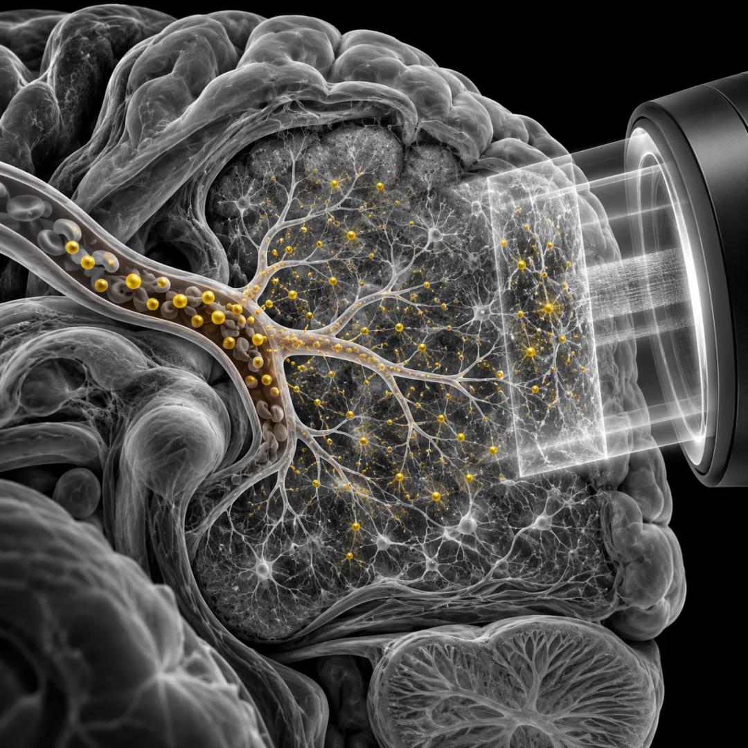
The oxygen extraction fraction is the share of the oxygen delivered to the brain by blood that tissue actually takes up. Raichle and colleagues used it in 2001 to give the brain a physiological baseline, and found that in people awake and resting with their eyes closed it was remarkably uniform across the brain. Regions whose oxygen use fell away from that baseline during goal-directed tasks became the first evidence for what is now called the default mode network.
What it is
Blood carries oxygen to the brain, and the tissue takes up some fraction of what arrives. That fraction is the oxygen extraction fraction, or OEF: the ratio of the oxygen the brain uses to the oxygen delivered by blood. It can be mapped with quantitative PET, which measures both the brain's oxygen metabolism and its blood flow, and the ratio follows from the two.
The measure matters because it ties the imaging signal to something physical. Functional MRI, the technique most readers have heard of, tracks local changes in the oxygen content of blood. Raichle and colleagues state in their abstract that local deviations in OEF underlie the signals seen with functional MRI during many human behaviours. A map of OEF is therefore a map of the thing that the more familiar scans are indirectly reading.
The 2001 paper, published in the Proceedings of the National Academy of Sciences, set out to define a baseline state of the normal adult human brain using this quantity. The choice of quantity was deliberate. A baseline defined by what a person is told to do, such as lie still and think of nothing, depends on what the person does with the instruction. A baseline defined by oxygen does not.
The vault records the use of OEF as a baseline as Raichle and colleagues' contribution. The measure itself is older, and this page does not fix its origin.
In effect
The finding that mattered was a negative one. When the authors mapped OEF throughout the brain in awake, resting people with their eyes closed, they found it remarkably uniform. In their words, areas of activation were conspicuous by their absence. The significant deviations from the baseline they did find were increases in OEF, which the authors interpreted as deactivations, and they lay almost entirely in the visual system.
From that baseline the paper turned to the areas that consistently decreased from it during goal-directed tasks. Those decreases were organised and recurring, and the authors proposed that they pointed to a default mode of brain function that is suspended during specific goal-directed behaviours. A later review by Raichle describes the network as a set of matched, bilateral regions in the parietal, medial prefrontal and temporal cortices, and says it was found largely by accident, in early PET studies that compared attention-demanding tasks with relaxed rest.
The practical effect for the argument in the article this page belongs to is on the objection that rest is not a proper reference condition. A baseline written in oxygen answers part of that objection before it is made, because nobody had to be told to think of nothing. It does not answer all of it, and the objection is set out on its own page.
What it does not say
It does not say what the default mode network is for. A uniform oxygen baseline and a set of regions that fall away from it during tasks establish that a pattern exists. They say nothing about what the pattern does.
It does not settle the objection raised by Alexa Morcom and Paul Fletcher in 2007, that activity at rest has no privileged status as a fundamental metric of brain function. A physiological definition makes the baseline less dependent on instructions. It does not show that a resting state is the right reference for every other state.
It does not link the network to mind-wandering. That link rests on later thought-sampling studies, and the field has not settled whether the default mode network is the mind-wandering network.
This page describes a measurement and a research finding. It carries no clinical guidance.
Sources
- Raichle, M. E., MacLeod, A. M., Snyder, A. Z., Powers, W. J., Gusnard, D. A., & Shulman, G. L. (2001). "A default mode of brain function." Proceedings of the National Academy of Sciences, 98(2), 676-682. doi:10.1073/pnas.98.2.676. Abstract read.
- Raichle, M. E. (2015). "The brain's default mode network." Annual Review of Neuroscience, 38, 433-447. doi:10.1146/annurev-neuro-071013-014030. Abstract read.
- Morcom, A. M., & Fletcher, P. C. (2007). "Does the brain have a baseline? Why we should be resisting a rest." NeuroImage, 37(4), 1073-1082. doi:10.1016/j.neuroimage.2006.09.013. Abstract read.